Digital Mole Scanning Clinic
If you are becoming increasingly concerned about a mole, we will analyse your moles with the best technology available giving you a sound diagnosis and peace of mind.
All of our treatments are carried out by qualified doctors and take place in our clinics located across South London. The locations include Clapham Junction (Northcote Road), Clapham North and Wandsworth.
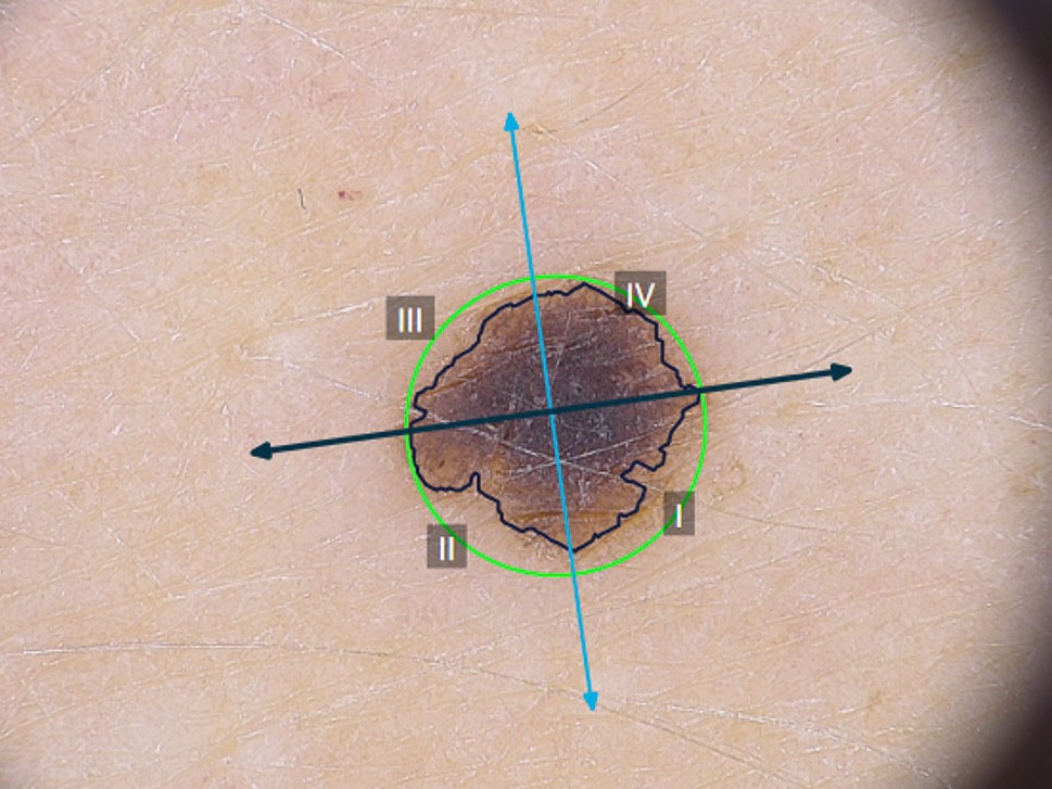
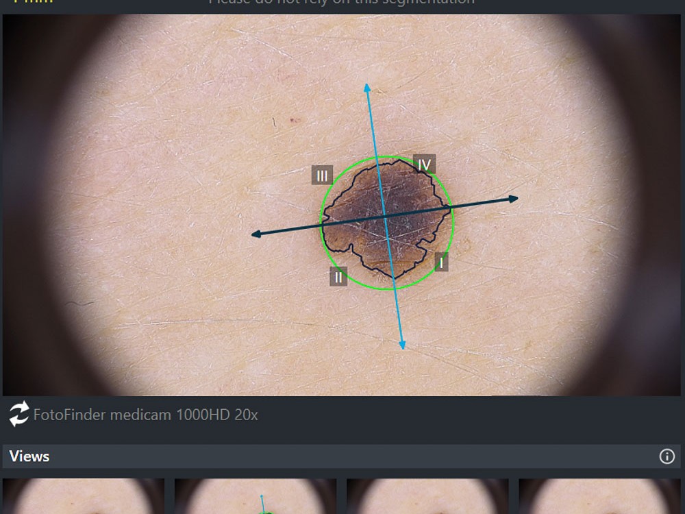
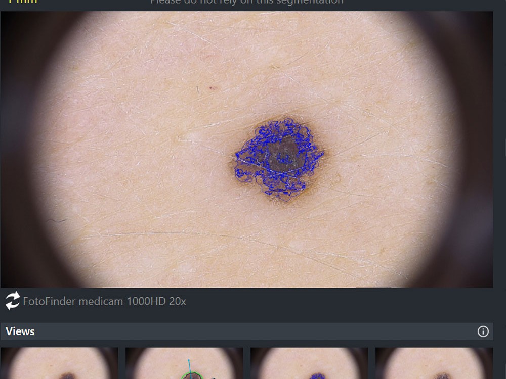
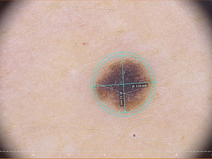
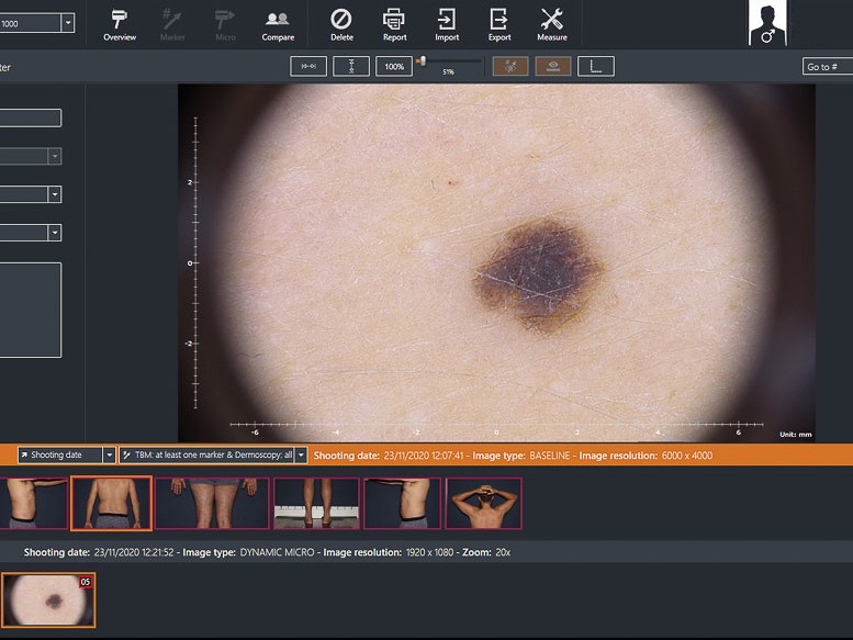
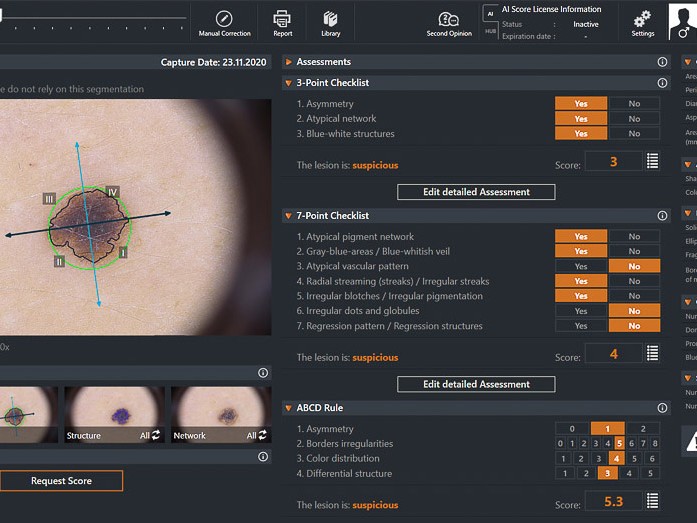
Example scan
Click to expand images
Digital Mole Scanning
EpicDermis featured on London LIVE
What is SIAscopy™?
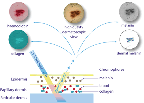
Spectrophotometric Intracutaneous Analysis (SIA) is a fast, non-invasive and completely safe method provides color bitmaps called SIAscans™ that show the relative location of blood, collagen and pigment. It is easy to perform and allows examination of skin lesions. SIAscopy™ understands the way that light interacts with the skin; the manner in which it scatters or bounces, the amount absorbed by cells and other structures along with the different changes in wavelength or color. The SIAscope™ provides color bitmaps called SIAscans™ that show the relative location of blood, collagen and pigment.
How does Siascopy™ work?
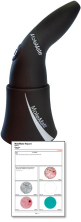
MoleMate™ and SIMSYS™ both use the Siascope™ handheld scanner. When the scanner illuminates the skin, some of the light is reflected and scattered from the surface. The remainder is transmitted into the top layers of the skin. Varying fractions of the incoming light are then firstly absorbed by the pigment in the epidermis before entering the dermis, where they are absorbed by the hemoglobin in the blood vessels. Scattering also occurs in the dermis when the light interacts with the collagen, resulting in a portion of the light being remitted back to the surface. By interpreting the combination of wavelengths that are received back by the SIAscope™ and is then able to produce SIAscans™; these are generated by referring to inbuilt proprietary mathematical models of skin optics. MoleMate™ is then able to present the user with the generated SIAscans™ for interpretation by the medical professional.
SKIN CANCER – Should I be worried?
Skin cancer is now the commonest form of cancer in the UK. The number of cases is increasing year after year.
Latest figures from the British Skin Foundation tell us that there are 100,000 new cases of skin cancer every single year. Sadly 2,500 people die from skin cancer every year. This is 2500 too many and our mole analysis clinic aims to prevent these sorts of diagnoses.
There are three main forms of skin cancer.
1. Basal cell carcinomas
2. Squamous cell carcinomas
3. Melanoma
Melanoma
Is the the melanoma type that is most difficult to spot, and the one that is most aggressive. Left undiagnosed and untreated – it can lead to widespread disease and the outcomes are usually very poor and sadly, often fatal. What often looks like an innocuous mole – often turns out to be a dangerous skin cancer. It can affect anyone – at ANY age.
Using our latest technology we can use cutting edge techniques to give us a unique insight into your moles and tell you if your mole has suspicious features or not.
As a doctor I see many skin cancers every year. The current methodology of reviewing skin cancer is based around a dated and non specific algorithm. We essentially just measure the size of the mole and monitor its shape, size, borders, colour and the part of the body that it is on. It’s known as the ABCDE rules. More on this later.
After this it’s watch and wait.
Here at EpicDermis we think we can do better than this. Better even than the current private mole mapping systems out there which essentially just take a series of high resolution photos and compare them over time. Our system is different. And unique. It uses the very latest technology to look underneath the skin up to depths of 4mm – something other clinics can’t currently do with their mole mapping.
If you can look underneath the skin, and see what feeds the mole, the blood supply, the pigmentation, and understand the mole, then our chances of identifying the cancerous mole from the innocent one have increased enormously.
Don’t risk it.
We analyse your moles with the best technology available giving you a sound diagnosis and peace of mind. In the unfortunate event when we think there is something suspicious about your mole then we can point you in the right direction to get it addressed and removed should you need.
All of our treatments are carried out by qualified doctors and take place in our clinics located across South London. The locations include Clapham Junction (Northcote Road), Clapham North, Earlsfield and Wandsworth. If you would like to arrange a free consultation please complete our enquiry form or alternatively email enquiries@epicdermis.co.uk.
The rates of melanoma are rising faster than any other type of cancer.
Why take the chance?
Frequently Asked Questions
1. What happens during the mole mapping or mole check?
During the mole mapping procedure (or Automated Total Body Mapping - ATBM), your entire body will be scanned and compared with a baseline image, to detect any visible marks and/or cancerous cells. The follow-up takes up to 45 minutes or so.
2.What happens if I need treatment?
In line with the mole mapping treatment, you will be asked to change to a medical gown. Following this, a specialized clinical computer will take scans of every inch of your body. The photos will be combined altogether to see the visible marks and/or moles.
3. When should you do mole mapping?
You should take up a mole mapping treatment if you have a high risk for skin cancer. This helps in the prevention of melanoma at the early stages itself.
4. Is mole mapping effective?
Yes, mole mapping is very much effective for detecting early-stage cancers, plus, for monitoring pigmented lesions. Hence, saving your cost, time, and life as well.
5. How do they test moles for skin cancer?
Skin cancer tests on moles are termed as “Biopsy”. A numbing shot is injected into the site (i.e. skin). The doctor will then try to scrape off the mole possibly from the skin. This is a 15 to 30-minute procedure.


